
Full resolution (JPEG) - On this page / på denna sida - Sidor ...
<< prev. page << föreg. sida << >> nästa sida >> next page >>
Below is the raw OCR text
from the above scanned image.
Do you see an error? Proofread the page now!
Här nedan syns maskintolkade texten från faksimilbilden ovan.
Ser du något fel? Korrekturläs sidan nu!
This page has never been proofread. / Denna sida har aldrig korrekturlästs.
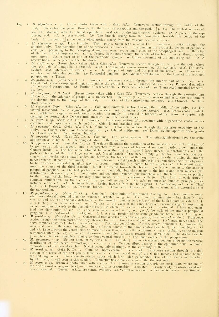
Fig. i. 31. giganteum, n. sp. (From photo, taken with a Zeiss AA.) Transvçrse section through the middle of the
body. The section has passed through the third pair of parapodia and the penis (^). b.s. The ventral nerve-cord.
mv. The stomach, with its ciliated epithelium, ov.d. One of the latero-ventral oviducts, s.h. A piece of the
supporting rod. r.li. A reserve-hook. li.k. The branch issuing from the hook-gland towards the centre of the
body. In the penis (j), the ductus ejaculatorius issuing from the vesicula seminalis is seen.
2. 31. giganteum, n. sp. Dwarf-male. (From photo, taken with a Zeiss CC.) Transverse section through the
anterior body. The posterior part of the proboscis is transsected. Surrounding the proboscis, groups of ganglionic
cells (gr.), pertaining to the oesophageal ring are seen. or. A small piece of the oesophageal ring. n. Branches
of the first pair of large nerves, t., t., t. Testes, distributed through the whole of the body. m.v.i. Musculi
retract-ores interni. f.g. A part of one of the parapodial ganglia, sh. Upper extremity of the supporting rod. r.li. A
reserve-hook. li. A piece of the chief-hook.
3. 31. graffi, 11. sp. (From photo, taken with a Zeiss AA.) Transverse section through the body, at the point where
the 4th pair of parapodia is situated, m.v.k. The glandulous mass surrounding the stomach, ov.d’. The dorsal
oviduct, ov.d. Latero-ventral oviduct. «.., n. The 5th pair of large, and 6th pair of fine nerves, dv. Dorso-ventral
muscles, mc. Musculus centralis, f.g. Parapodial ganglion, p.f. Annular protuberance at the base of the retracted
parapodium. t. Testes.
» 4. 31. graffi, n. sp. (Zeiss AA. Oc. 1. Cam.-luc.) Transverse section through the anterior part of the body. 0. r.
Dorsal part of the oesophageal ring, surrounding the proboscis, n., n. Transsected nerves, f.g. Parapodial ganglion
of the second parapodium. r.li. Portion of reserve-hook. h. Piece of chief-hook. tvi. Transsected intestinal branehes.
ov, Ova.
31. cirriferum, F. S. Leuck. (From photo, taken with a Zeiss CC.) Transverse section through the posterior part
of the body; the 4th pair of parapodia is transsected. The testes (t., t., t.) are seen extending to the ectoderm of
the dorsum and to the margin of the body. ov.d. One of the ventro-lateral oviducts, m.v. Stomach, tm.
Intestinal branches.
» 6. 31. carpenteri, Graff. (Zeiss AA. Oc. 1. Cam.-luc.)Transverse section through the middle of the body. b.s. The
ventral nerve-cord. s.o. A segmental glandulous sack. spli. Sphincter of the aperture, ov.d. Latero-ventral oviduct.
ov.r. Problematic organs — rudimentary ovaries presumably — situated in branches of the uterus, d. Septum sub
dividing the uterus, d. v. Dorso-ventral muscles, dr. The dorsal ridges.
7. 31. graffi, n. sp. (Zeiss AA. Oc. 1. Cam.-luc.) Transverse section of a specimen with degenerated ventral
nerve-cord (b.s.), and capacious stomach (mv.) from which lateral branches issue.
8. 31. carpenteri, Graff. (Zeiss AA. Oc. 1. Cam.-luc.) Transverse section through the posterior extremity of the
body. ei. Cloacal canal, an. Cloacal aperture, f.e. Ciliated epithelium, ovd. Dorsal oviduct-aperture opening into
the cloacal aperture, tm. Intestinal branches.
9. 31. carpenteri, Grrff. (Zeiss CC. Oc. 2. Cam.-luc.) The cloacal aperture. The leitra-significations have the same
application as in fig. 8. f. The tongued labiæ of the aperture.
» 10. 31. giganteum, n. sp. (Zeiss AA. Oc. 5.) The figure illustrates the distribution of the sinistral nerve of the first pair of
large nerves (dorsal aspect), and is constructed from a series of horizontal sections; partly, drawn under the
Camera lucida, n. The fine nerve situated between the first and second large nerves; it anastomozes with the
posterior branch of the first large nerve, n.’. Mesial branch of the nerve ramifying into two branchlets, one
passing to the muscles (m.) situated under, and between, the branches of the large nerve; the other crossing the anterior
nerve-branches; it passes, presumably, to the muscles m.’. n? A branch ramifying into 3 branchlets, one of which passes
to be posterior parapodial ganglion (f.g.), a second branchlet (n3) passes to the muscles m.3. I have not
exam-imed the course of the third branchlet, but it is directed towards the hooks, g.n. Nerve running to the anterior
parapodial ganglion, d. The first portion of nerve (parapodial branch) running to the hooks and their muscles (the
distribution is shown in fig. 11). The anterior and posterior branches (cirri-branches), are, the large branches passing
to the margin of the body, where they communicate with the epidermic cells (b.). They form, at each cirrus, a
complex ramification. In their course, they give off nerves (m.4, n.b, rø,6), to muscular bundles principally (for
instance, n.6 to m,2). h.k. The inner branch, issuing inwards from the hook-gland, li. Supporting rod. s. li. Chief
hook. r. li. Reserve-hook. tm. Intestinal branch, c. Transsected depression in the ventrum, at the external side of
the parapodium.
» 11. 31. giganteum, n. sp. (Zeiss CC. Oc. 4. Cam.-luc.) Distribution of the branch d of fig. 10. This branch is
somewhat more dorsally situated than the branches illustrated in fig. 10. The branch ramifies into 3 branchlets (n.’, n",
n.3), and n.2, are principally distributed in the muscular bundles (m m.-, m3.) of the hook-apparatus, vide 1, 2, 3,
4, 5, 6 etc.; some branchlets (n.", and n’") pass to the walls of the canal however, encompassing the supporting
rod (h.), and pass onwards to the glandular mass (a.) in which the reserve hooks (r.li.) are situated. I have not
examined the distribution of n.3. n.4 is the same nerve as w.6 in fig. 10. f.g. A few cells of the anterior parapodial
ganglion, b. A portion of the hook-gland, li. lc. A small portion of the same glandulous branch as h k. in fig. 10.
» 12. 31. graffi, n. sp. (Zeiss AA. Oc. 1. Constructed from a series of sections and, partly, drawn under Cam.-luc.) Transverse
scction through the mesial part of the bod)’, showing the distribution of one of the fine nerves, b.s. Ventral nerve-cord. The
nerve ramifies at its root into two branches (2, 3). From the ventral one, of these, several branchlets (1), immediately,
issue and pass to the ventral muscles. In the further course of the same ventral branch (2), the branchlets n.’, ws
and n.3, issue towards the ventral side, to muscles as well as, also, to the ectoderm; n.* runs, probably, to the musculi
retractores interni (m. r.)\ m.5, runs to dorso-ventral muscles; 4 passes towards the dorsal side. The dorsal branch,
3, ramifies into two branchlets running to dorso-ventral muscles, f. The inner outline of the parapodium.
» 13. 31. giganteum, n. sp. (Seibert hom. im ’/s Zeiss Oc. 1. Cam.-luc.) From a transverse section, showing the vertical
distribution of the nerve terminating in a cirrus, a., a. Nervous fibres passing to the epidermic cells, b.
Anas-tomozations of the nerve-branches. Nuclei occur, only sparingly, at the extremity of the cirrus.
» 14. 31. graffi, n sp. (Zeiss F. Oc. 1. Cam.-luc. Borax-carmine. Picric acid.) Transverse section through the first
portion of a nerve of the first pair of large nerves, n. The second one of the 6 fine nerves, and situated behind
the first large nerve. The connective-tissue septa which form »den gefacherten Bau« of the nerves, as described
by Hermann, is well seen in this section. Connective-tissue nuclei occur in the thickest septa.
» 15. 31. graffi, n. sp. (From a photo taken with a Zeiss CC.) Transverse section through the mesial part, where one of
the problematic organs, (ovr.), — rudimentary ovaries presumably — is situated, a. Body-cavity, 011 whose dorsal side
ova are situated, t. Testes, ovd. Latero-ventral oviducts, b.s. Ventral nerve-cord. n. Transsected nerve, mv. Stomach.
<< prev. page << föreg. sida << >> nästa sida >> next page >>