
Full resolution (JPEG) - On this page / på denna sida - Sidor ...
<< prev. page << föreg. sida << >> nästa sida >> next page >>
Below is the raw OCR text
from the above scanned image.
Do you see an error? Proofread the page now!
Här nedan syns maskintolkade texten från faksimilbilden ovan.
Ser du något fel? Korrekturläs sidan nu!
This page has never been proofread. / Denna sida har aldrig korrekturlästs.
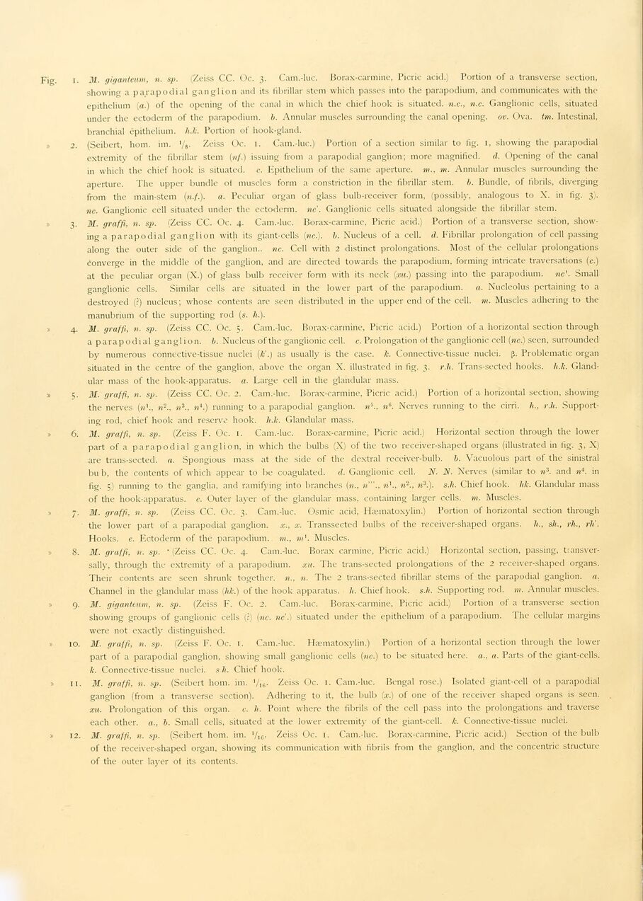
Fig. i. 31. giganteum, n. sp. Zeiss CC. Oc. 3. Cam.-luc. Borax-carmine, Picric acid.) Portion of a transverse section,
showing a parapodial ganglion and its fibrillar stem which passes into the parapodium, and communicates with the
epithelium (a.) of the opening of the canal in which the chief hook is situated, n.c., n.c. Ganglionic cells, situated
under the ectoderm of the parapodium. b. Annular muscles surrounding the canal opening, ov. Ova. tun. Intestinal,
branchial epithelium, h.k. Portion of hook-gland.
2. (Seibert, hom. im. ’/a• Zeiss Oc. 1. Cam.-luc.) Portion of a section similar to fig. 1, showing the parapodial
extremity of the fibrillar stem (nf.) issuing from a parapodial ganglion; more magnified. <1. Opening of the canal
in which the chief hook is situated, c. Epithelium of the same aperture. m., m. Annular muscles surrounding the
aperture. The upper bundle of muscles form a constriction in the fibrillar stem. b. Bundle, of fibrils, diverging
from the main-stem («./.). a. Peculiar organ of glass bulb-receiver form, (possibly, analogous to X. in fig. 3).
nc. Ganglionic cell situated under the ectoderm, nc. Ganglionic cells situated alongside the fibrillar stem.
3. ill. graffi, n. sj). (Zeiss CC. Oc. 4. Cam.-luc. Borax-carmine, Picric acid.) Portion of a transverse section,
showing a parapodial ganglion with its giant-cells (nc.). b. Nucleus of a cell. d. Fibrillar prolongation of cell passing
along the outer side of the ganglion., nc. Cell with 2 distinct prolongations. Most of the cellular prolongations
Converge in the middle of the ganglion, and are directed towards the parapodium, forming intricate traversations (c.)
at the peculiar organ (X.) of glass bulb receiver form with its neck (xu.) passing into the parapodium. ne’. Small
ganglionic cells. Similar cells are situated in the lower part of the parapodium. a. Nucleolus pertaining to a
destroyed (?) nucleus; whose contents are seen distributed in the upper end of the cell. m. Muscles adhering to the
manubrium of the supporting rod (s. /».).
4. 31. graffi, n. sp. (Zeiss CC. Oc. 5. Cam.-luc. Borax-carmine, Picric acid.) Portion of a horizontal section through
a parapodial ganglion, b. Nucleus of the ganglionic cell. c. Prolongation of the ganglionic cell (nc.) seen, surrounded
by numerous connc-ctive-tissue nuclei (k’.) as usually is the case. k. Connective-tissue nuclei, ß. Problematic organ
situated in the centre of the ganglion, above the organ X. illustrated in fig. 3. r.h. Trans-sected hooks, h.k.
Glandular mass of the hook-apparatus, a. Large cell in the glandular mass.
» 5. 31. graffi, n. sp. (Zeiss CC. Oc. 2. Cam.-luc. Borax-carmine, Picric acid.) Portion of a horizontal section, showing
the nerves («’., n-., n3., n4.) running to a parapodial ganglion, n’1., n6. Nerves running to the cirri. /<., r.h.
Supporting rod, chief hook and reserve hook. h.k. Glandular mass.
6. M. graffi, n. sp. (Zeiss I7. Oc. 1. Cam.-luc. Borax-carmine, Picric acid.) Horizontal section through the lower
part of a parapodial ganglion, in which the bulbs (X) of the two receiver-shaped organs (illustrated in fig. 3, X)
are trans-sected. a. Spongious mass at the side of the dextral receiver-bulb. b. Vacuolous part of the sinistral
bu b, the contents of which appear to be coagulated, d. Ganglionic cell. N. N. Nerves (similar to n3. and n4. in
fig. 5) running to the ganglia, and ramifying into branches (n., n’"., n\, n-., n3.). s.h. Chief hook. lik. Glandular mass
of the hook-apparatus, c. Outer layer of the glandular mass, containing larger cells, m. Muscles.
7. 31. graffi, n. sp. (Zeiss CC. Oc. 3. Cam.-luc. Osmic acid, Hæmatoxylin.) Portion of horizontal section through
the lower part of a parapodial ganglion, x., x. Transsected bulbs of the receiver-shaped organs, h., sh., rh., rli.
Hooks, e. Ectoderm of the parapodium. m., w’. Muscles.
8. 31. graffi, n. sp. " (Zeiss CC. Oc. 4. Cam.-luc. Borax carmine, Picric acid.) Horizontal section, passing,
tiansver-sally, through the extremity of a parapodium. xu. The trans-sected prolongations of the 2 receiver-shaped organs.
Their contents are seen shrunk together. n., n. The 2 trans-sected fibrillar stems of the parapodial ganglion, a.
Channel in the glandular mass (lik.) of the hook apparatus, li. Chief hook. s.h. Supporting rod. m. Annular muscles.
9. 31. giganteum, n. sp. (Zeiss F. Oc. 2. Cam.-luc. Borax-carmine, Picric acid.) Portion of a transverse section
showing groups of ganglionic cells (?) (nc. ne’.) situated under the epithelium of a parapodium. The cellular margins
were not exactly distinguished.
» 10. M. graffi, n. sp. (Zeiss F. Oc. 1. Cam.-luc. Hæmatoxylin.) Portion of a horizontal section through the lower
part of a parapodial ganglion, showing small ganglionic cells (nc.) to be situated here, a., a. Parts of the giant-cells.
k. Connective-tissue nuclei, s h. Chief hook.
» 11. 31. graffi, n. sp. (Seibert hom. im. ’/16. Zeiss Oc. 1. Cam.-luc. Bengal rose.) Isolated giant-cell of a parapodial
ganglion (from a transverse section1). Adhering to it, the bulb (x.) of one of the receiver shaped organs is seen.
xu. Prolongation of this organ, c. h. Point where the fibrils of the cell pass into the prolongations and traverse
each other, a., b. Small cells, situated at the lower extremity of the giant-cell. k. Connective-tissue nuclei.
» 12. 31. graffi, n. sp. (Seibert hom. im. ’/IC. Zeiss Oc. 1. Cam.-luc. Borax-carmine, Picric acid.) Section of the bulb
of the receiver-shaped organ, showing its communication with fibrils from the ganglion, and the concentric structure
of the outer layer of its contents.
<< prev. page << föreg. sida << >> nästa sida >> next page >>