
Full resolution (JPEG) - On this page / på denna sida - Sidor ...
<< prev. page << föreg. sida << >> nästa sida >> next page >>
Below is the raw OCR text
from the above scanned image.
Do you see an error? Proofread the page now!
Här nedan syns maskintolkade texten från faksimilbilden ovan.
Ser du något fel? Korrekturläs sidan nu!
This page has never been proofread. / Denna sida har aldrig korrekturlästs.
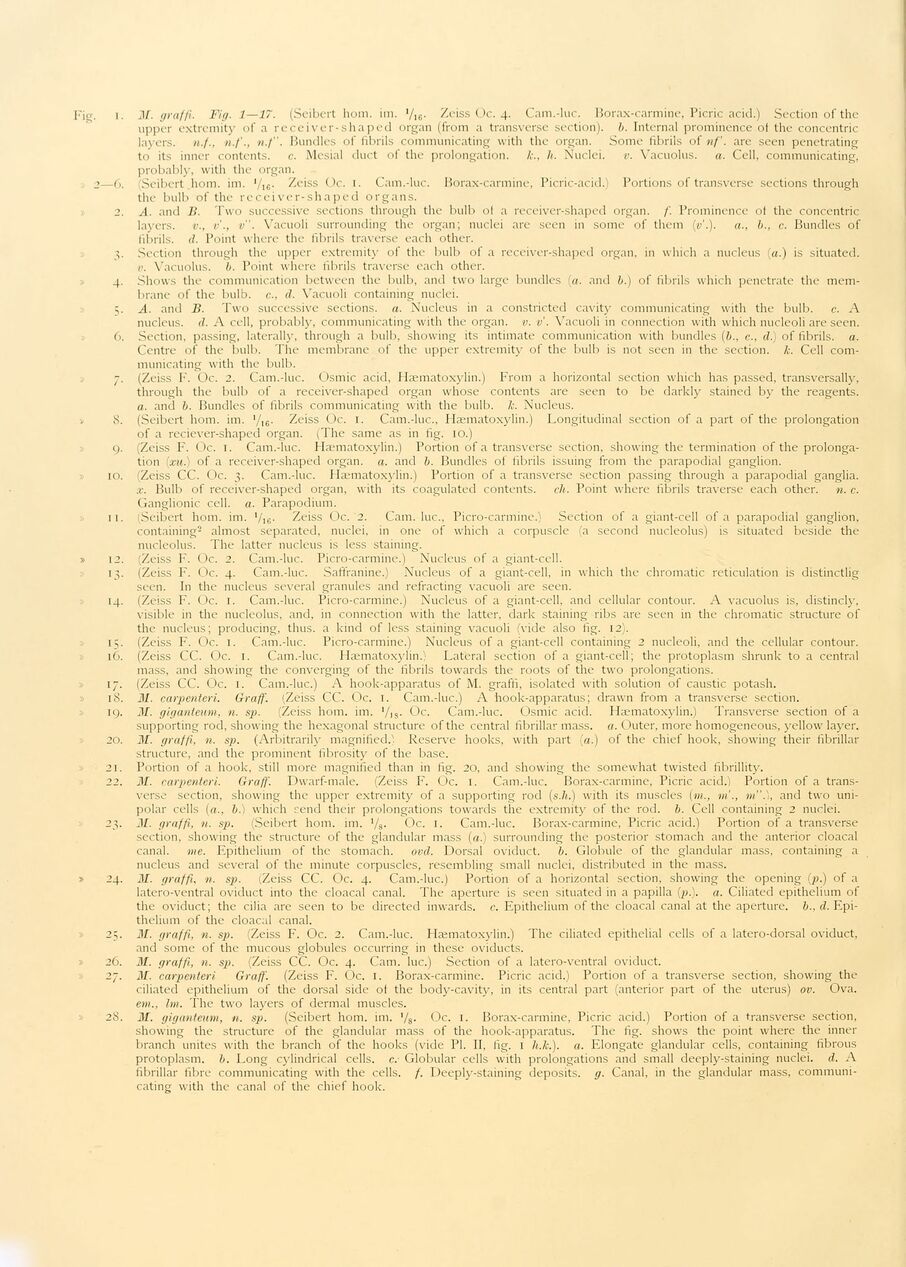
Fig. i. M. graffi. Fig. 1—17. (Seibert hom. im. Vie- Zeiss Oc. 4. Cam.-lue. Borax-cärmine, Picric acid.) Section of the
upper extremity of a receiver-shaped organ (from a transverse section), b. Internal prominence ot the concentric
layers. n.f., n.f., n.f". Bundles of fibrils communicating with the organ. Some fibrils of nf. are seen penetrating
to its inner contents, c. Mesial duct of the prolongation, k., h. Nuclei. v. Vacuolus. a. Cell, communicating,
probably, with the organ.
2—6. Seibert hom. im. ’/16. Zeiss Oc. 1. Cam.-luc. Borax-carmine, Picric-acid.) Portions of transverse sections through
the bulb of the receiver-shaped organs.
2. A. and B. Two successive sections through the bulb of a receiver-shaped organ. /’. Prominence ol the concentric
layers, v., v., v". Vacuoli surrounding the organ; nuclei are seen in some of them (v’.). a., b., c. Bundles of
fibrils, d. Point where the fibrils traverse each other.
Section through the upper extremity of the bulb of a receiver-shaped organ, in which a nucleus (a.) is situated.
v. Vacuolus. b. Point where fibrils traverse each other.
4. Shows the communication between the bulb, and two large bundles (a. and b.) of fibrils which penetrate the
membrane of the bulb, c., d. Vacuoli containing nuclei.
5. A. and B. Two successive sections, a. Nucleus in a constricted cavity communicating with the bulb. c. A
nucleus, ei. A cell, probably, communicating with the organ, v. v. Vacuoli in connection with which nucleoli are seen.
6. Section, passing, laterally, through a bulb, showing its intimate communication with bundles (&., c., d.) of fibrils, a.
Centre of the bulb. The membrane of the upper extremity of the bulb is not seen in the section, k. Cell
communicating with the bulb.
7. (Zeiss F. Oc. 2. Cam.-luc. Osmic acid, Hæmatoxylin.) From a horizontal section which has passed, transversally,
through the bulb of a receiver-shaped organ whose contents are seen to be darkly stained by the reagents.
a. and b. Bundles of fibrils communicating with the bulb. k. Nucleus.
» 8. (Seibert hom. im. ’/,6. Zeiss Oc. 1. Cam.-luc., Hæmatoxylin.) Longitudinal section of a part of the prolongation
of a reciever-shaped organ. (The same as in fig. 10.)
9. ;Zeiss F. Oc. 1. Cam.-luc. Hæmatoxylin.) Portion of a transverse section, showing the termination of the
prolongation (xu.) of a receiver-shaped organ, a. and b. Bundles of fibrils issuing from the parapodial ganglion.
10. Zeiss CC. Oc. 3. Cam.-luc. Hæmatoxylin.) Portion of a transverse section passing through a parapodial ganglia.
x. Bulb of receiver-shaped organ, with its coagulated contents, ch. Point where fibrils traverse each other, n. c.
Ganglionic cell. a. Parapodium.
11. (Seibert hom. im. Vie- Zeiss Oc. 2. Cam. luc., Picro-carmine.) Section of a giant-cell of a parapodial ganglion,
containing2 almost separated, nuclei, in one of which a corpuscle (a second nucleolus) is situated beside the
nucleolus. The latter nucleus is less staining.
» 12. (Zeiss F. Oc. 2. Cam.-luc. Picro-carmine.) Nucleus of a giant-cell.
13. (Zeiss F. Oc. 4. Cam.-luc. Satfranine.) Nucleus of a giant-cell, in which the chromatic reticulation is distinctlig
seen. In the nucleus several granules and refracting vacuoli are seen.
14. (Zeiss F. Oc. 1. Cam.-luc. Picro-carmine.) Nucleus of a giant-cell, and cellular contour. A vacuolus is, distincly,
visible in the nucleolus, and, in connection with the latter, dark staining ribs are seen in the chromatic structure of
the nucleus; producing, thus, a kind of less staining vacuoli (vide also fig. 12).
15. (Zeiss F. Oc. 1. Cam.-luc. Picro-carmine.) Nucleus of a giant-cell containing 2 nucleoli, and the cellular contour.
16. (Zeiss CC. Oc. 1. Cam.-luc. Hæmatoxylin.) Lateral section of a giant-cell; the protoplasm shrunk to a central
mass, and showing the converging of the fibrils towards the roots of the two prolongations.
17. (Zeiss CC. Oc. 1. Cam.-luc.) A hook-apparatus of M. graffi, isolated with solution of caustic potash.
18. M. carpenteri. Graff. (Zeiss CC. Oc. 1. Cam.-luc.) A hook-apparatus; drawn from a transverse section.
19. M. giganteum. n. sp. (Zeiss hom. im. ’/is- Oc. Cam.-luc. Osmic acid. Hæmatoxylin.) Transverse section of a
supporting rod, showing the hexagonal structure of the central fibrillar mass. a. Outer, more homogeneous, yellow layer.
20. M. graffi, n. sp. (Arbitrarily magnified.) Reserve hooks, with part (a.) of the chief hook, showing their fibrillar
structure, and the prominent fibrosity of the base.
21. Portion of a hook, still more magnified than in fig. 20, and showing the somewhat twisted fibrillity.
22. M. carpenteri. Graff. Dwarf-male. (Zeiss F. Oc. 1. Cam.-luc. Borax-carmine, Picric acid.) Portion of a
transverse section, showing the upper extremity of a supporting rod (s.h.) with its muscles (m., m’., m". 1, and two
unipolar cells (a., b.) which rend their prolongations towards the extremity of the rod. b. Cell containing 2 nuclei.
23. 31. graffi, n. sp. (Seibert hom. im. 1fs. Oc. 1. Cam.-luc. Borax-carmine, Picric acid.) Portion of a transverse
section, showing the structure of the glandular mass (a.) surrounding the posterior stomach and the anterior cloacal
canal, me. Epithelium of the stomach. ovd. Dorsal oviduct. b. Globule of the glandular mass, containing a
nucleus and several of the minute corpuscles, resembling small nuclei, distributed in the mass.
» 24. M. graffi. n. sp. (Zeiss CC. Oc. 4. Cam.-luc.) Portion of a horizontal section, showing the opening (p.) of a
latero-ventral oviduct into the cloacal canal. The aperture is seen situated in a papilla (p.). a. Ciliated epithelium of
the oviduct; the cilia are seen to be directed inwards, c. Epithelium of the cloacal canal at the aperture, b., d.
Epithelium of the cloacal canal.
25. M. graffi, n. sp. (Zeiss F. Oc. 2. Cam.-luc. Hæmatoxylin.) The ciliated epithelial cells of a latero-dorsal oviduct,
and some of the mucous globules occurring in these oviducts.
» 26. M. graffi, n. sp. (Zeiss CC. Oc. 4. Cam. luc.) Section of a latero-ventral oviduct.
27. M. carpenteri Graff. (Zeiss F. Oc. 1. Borax-carmine. Picric acid.) Portion of a transverse section, showing the
ciliated epithelium of the dorsal side of the body-cavity, in its central part (anterior part of the uterus) ov. Ova.
em., Im. The two layers of dermal muscles.
28. M. giganteum, n. sp. (Seibert hom. im. */s- Oc. 1. Borax-carmine, Picric acid.) Portion of a transverse section,
showing the structure of the glandular mass of the hook-apparatus. The fig. shows the point where the inner
branch unites with the branch of the hooks (vide Pl. II, fig. 1 h.k.). a. Elongate glandular cells, containing fibrous
protoplasm, b. Long cylindrical cells, c. Globular cells with prolongations and small deeply-staining nuclei, d. A
fibrillar fibre communicating with the cells, f. Deeply-staining deposits, g. Canal, in the glandular mass,
communicating with the canal of the chief hook.
<< prev. page << föreg. sida << >> nästa sida >> next page >>