Full resolution (JPEG) - On this page / på denna sida - Sidor ...

<< prev. page << föreg. sida << >> nästa sida >> next page >>
Below is the raw OCR text
from the above scanned image.
Do you see an error? Proofread the page now!
Här nedan syns maskintolkade texten från faksimilbilden ovan.
Ser du något fel? Korrekturläs sidan nu!
This page has never been proofread. / Denna sida har aldrig korrekturlästs.
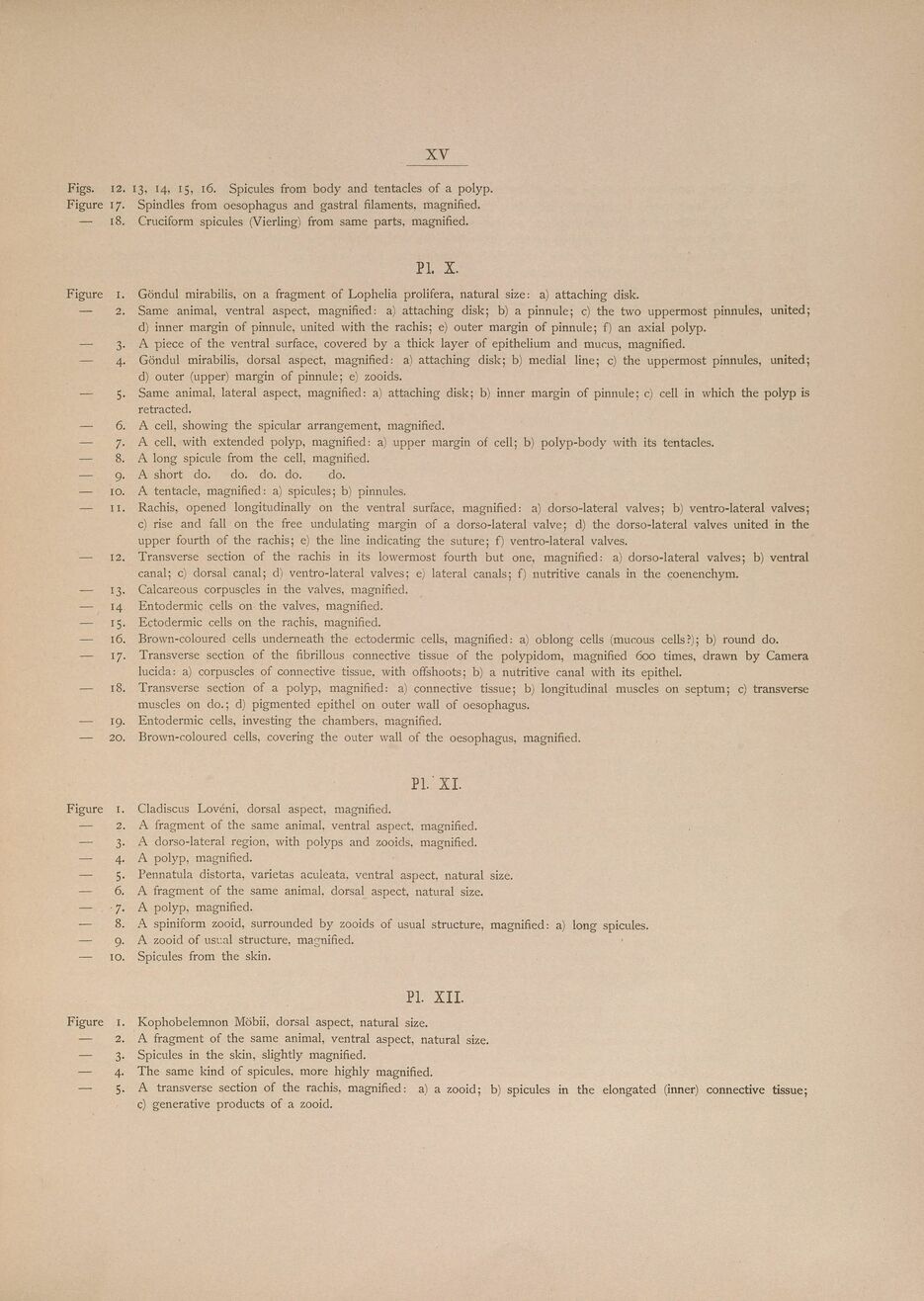
XV
Figs. 12. 13, 14, 15, 16. Spicules from body and tentacles of a polyp.
Figure 17. Spindles from oesophagus and gastral filaments, magnified.
— 18. Cruciform spicules (Vierling) from same parts, magnified.
Pl. X.
Figure 1. Göndul mirabilis, on a fragment of Lophelia prolifera, natural size: a) attaching disk.
— 2. Same animal, ventral aspect, magnified: a) attaching disk; b) a pinnule; c) the two uppermost pinnules, united;
d) inner margin of pinnule, united with the rachis; e) outer margin of pinnule; f) an axial polyp.
— 3. A piece of the ventral surface, covered by a thick layer of epithelium and mucus, magnified.
— 4. Göndul mirabilis, dorsal aspect, magnified: a) attaching disk; b) medial line; c) the uppermost pinnules, united;
d) outer (upper) margin of pinnule; e) zooids.
— 5. Same animal, lateral aspect, magnified: a) attaching disk; b) inner margin of pinnule; c) cell in which the polyp is
retracted.
— 6. A cell, showing the spicular arrangement, magnified.
— 7. A cell, with extended polyp, magnified: a) upper margin of cell; b) polyp-body with its tentacles.
— 8. A long spicule from the cell, magnified.
— 9. A short do. do. do. do. do.
— 10. A tentacle, magnified: a) spicules; b) pinnules.
— II. Rachis, opened longitudinally on the ventral surface, magnified: a) dorso-lateral valves; b) ventro-lateral valves;
c) rise and fall on the free undulating margin of a dorso-lateral valve; d) the dorso-lateral valves united in the
upper fourth of the rachis; e) the line indicating the suture; f) ventro-lateral valves.
— 12. Transverse section of the rachis in its lowermost fourth but one, magnified: a) dorso-lateral valves; b) ventral
canal; c) dorsal canal; d) ventro-lateral valves; e) lateral canals; f) nutritive canals in the çoenenchym.
— 13. Calcareous corpuscles in the valves, magnified.
— 14 Entodermic cells on the valves, magnified.
— 15. Ectodermic cells on the rachis, magnified.
— 16. Brown-coloured cells underneath the ectodermic cells, magnified: a) oblong cells (mucous cells?); b) round do.
— 17. Transverse section of the fibrillous connective tissue of the polypidom, magnified 600 times, drawn by Camera
lucida: a) corpuscles of connective tissue, with offshoots; b) a nutritive canal with its epithel.
— 18. Transverse section of a polyp, magnified: a) connective tissue; b) longitudinal muscles on septum; c) transverse
muscles on do.; d) pigmented epithel on outer wall of oesophagus.
— 19. Entodermic cells, investing the chambers, magnified.
— 20. Brown-coloured cells, covering the outer wall of the oesophagus, magnified.
Pl. ’XI.
Figure i. Cladiscus Lovéni, dorsal aspect, magnified.
— 2. A fragment of the same animal, ventral aspect, magnified.
— 3. A dorso-lateral region, with polyps and zooids, magnified.
— 4. A polyp, magnified.
— 5. Pennatula distorta, varietas aculeata, ventral aspect, natural size.
— 6. A fragment of the same animal, dorsal aspect, natural size.
— 7. A polyp, magnified.
— 8. A spiniform zooid, surrounded by zooids of usual structure, magnified: a) long spicules.
— 9. A zooid of usual structure, magnified.
— 10. Spicules from the skin.
Pl. XII.
Figure 1. Kophobelemnon Möbii, dorsal aspect, natural size.
— 2. A fragment of the same animal, ventral aspect, natural size.
— 3. Spicules in the skin, slightly magnified.
— 4. The same kind of spicules, more highly magnified.
— 5. A transverse section of the rachis, magnified: a) a zooid; b) spicules in the elongated (inner) connective tissue;
c) generative products of a zooid.
<< prev. page << föreg. sida << >> nästa sida >> next page >>