
Full resolution (JPEG) - On this page / på denna sida - Sidor ...
<< prev. page << föreg. sida << >> nästa sida >> next page >>
Below is the raw OCR text
from the above scanned image.
Do you see an error? Proofread the page now!
Här nedan syns maskintolkade texten från faksimilbilden ovan.
Ser du något fel? Korrekturläs sidan nu!
This page has never been proofread. / Denna sida har aldrig korrekturlästs.
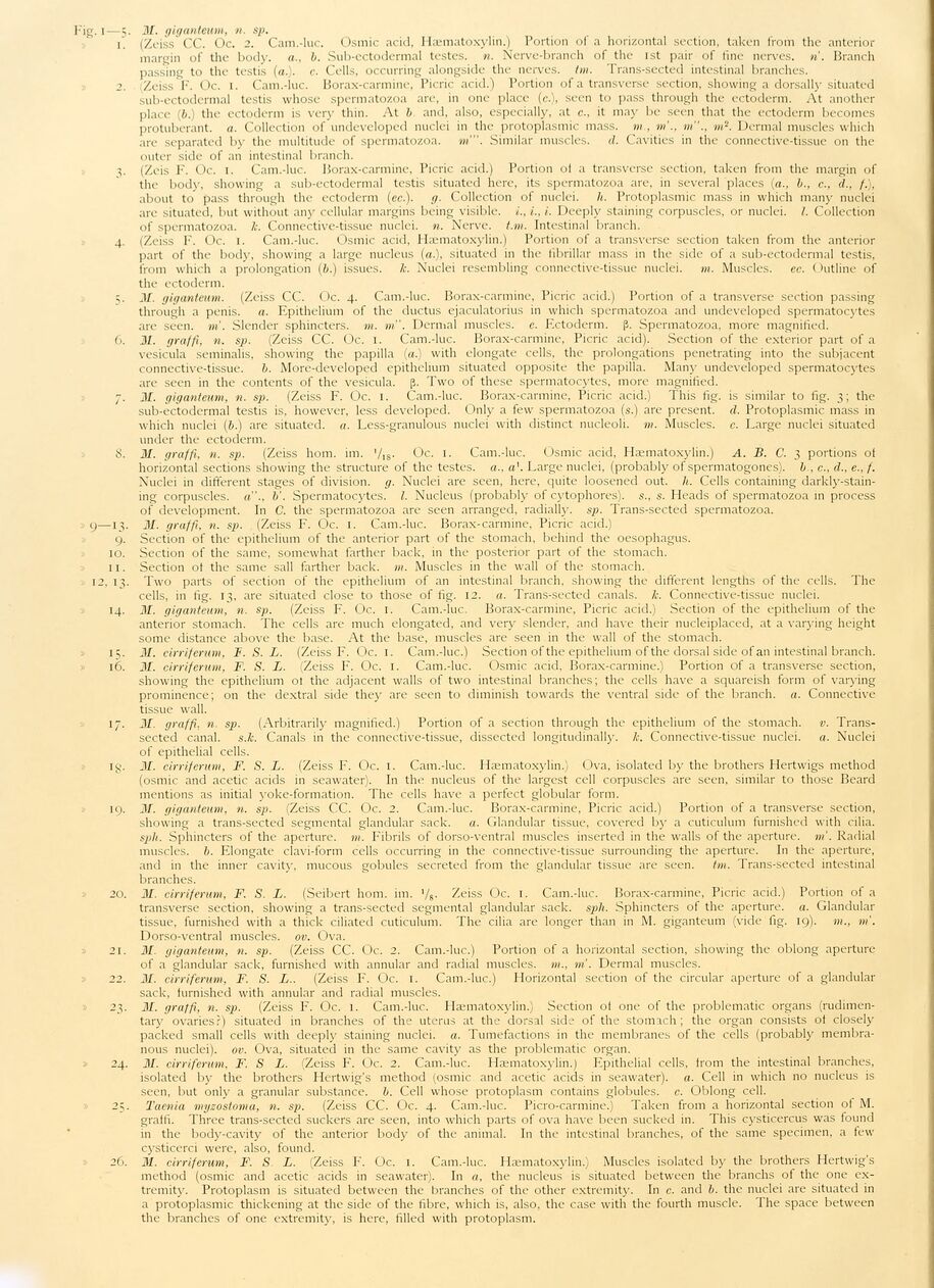
Fig. i—5. 31. giganteum, n. sp.
i . (Zeiss CC. Oc. 2. Cam.-luc. Osmic acid, Hæmatoxylin.) Portion of a horizontal section, taken from the anterior
margin of the bod)-, a., b. Sub-ectodermal testes, n. Nerve-branch of the lst pair of fine nerves, n’. Branch
passing to the testis (a.), c. Cells, occurring alongside the nerves, tm. Trans-sected intestinal branches.
2. Zeiss F. Oc. 1. Cam.-luc. Borax-carmine, Picric acid.) Portion of a transverse section, showing a dorsally situated
sub-ectodermal testis whose spermatozoa are, in one place (c.), seen to pass through the ectoderm. At another
place (6.) the ectoderm is very thin. At b and, also, especially, at c., it may be seen that the ectoderm becomes
protuberant, a. Collection of undeveloped nuclei in the protoplasmic mass, m , m’., m"., m’2. Dermal muscles which
are separated by the multitude of spermatozoa, m’". Similar muscles, d. Cavities in the connective-tissue on the
outer side of an intestinal branch.
i,. (Zeis F. Oc. I. Cam.-luc. Borax-carmine, Picric acid.) Portion ot a transverse section, taken from the margin of
the body, showing a sub-ectodermal testis situated here, its spermatozoa are, in several places (a., b., c., d., /.),
about to pass through the ectoderm (ec.). g. Collection of nuclei, h. Protoplasmic mass in which many nuclei
are situated, but without any cellular margins being visible, i., L, i. Deeply staining corpuscles, or nuclei. I. Collection
of spermatozoa. k. Connective-tissue nuclei, n. Nerve, t.m. Intestinal branch.
4. (Zeiss F. Oc. 1. Cam.-luc. Osmic acid, Hæmatoxylin.) Portion of a transverse section taken from the anterior
part of the body, showing a large nucleus (a.), situated in the fibrillar mass 111 the side of a sub-ectodermal testis,
from which a prolongation (b.) issues, lc. Nuclei resembling connective-tissue nuclei, tn. Muscles. ec. Outline of
the ectoderm.
5. 31. giganteum. (Zeiss CC. Oc. 4. Cam.-luc. Borax-carmine, Picric acid.) Portion of a transverse section passing
through a penis, a. Epithelium of the ductus ejaculatorius in which spermatozoa and undeveloped spermatocytes
are seen. m’. Slender sphincters, m. m’. Dermal muscles, c. Ectoderm. (J. Spermatozoa, more magnified.
6. 31. graffi, n. sp. (Zeiss CC. Oc. 1. Cam.-luc. Borax-carmine, Picric acid). Section of the exterior part of a
vesicula seminalis, showing the papilla (a.) with elongate cells, the prolongations penetrating into the subjacent
connective-tissue. b. More-developed epithelium situated opposite the papilla. Many undeveloped spermatocytes
are seen in the contents of the vesicula. ß. Two of these spermatocytes, more magnified.
7. 31. giganteum, n. sp. (Zeiss F. Oc. 1. Cam.-luc. Borax-carmine, Picric acid.) This fig. is similar to fig. 3; the
sub-ectodermal testis is, however, less developed. Only a few spermatozoa (s.) are present, d. Protoplasmic mass in
which nuclei (6.) are situated, a. Less-granulous nuclei with distinct nucleoli, m. Muscles, c. Large nuclei situated
under the ectoderm.
S. 31. graffi, n. sp. (Zeiss hom. im. ’/is- Oc. 1. Cam.-luc. Osmic acid, Hæmatoxylin.) A. B. C. 3 portions of
horizontal sections showing the structure of the testes, a., a\ Large nuclei, (probably of spermatogones), b , c., d., e., f.
Nuclei in different stages of division, g. Nuclei are seen, here, quite loosened out. h. Cells containing
darkly-staining corpuscles, a"., b’. Spermatocytes. I. Nucleus (probably of cytophores). s., s. Heads of spermatozoa in process
of development. In C. the spermatozoa are seen arranged, radially, sp. Trans-sected spermatozoa.
9—13. 31. graffi, n. sp. (Zeiss F. Oc. 1. Cam.-luc. Borax-carmine, Picric acid.)
9. Section of the epithelium of the anterior part of the stomach, behind the oesophagus.
10. Section of the same, somewhat farther back, in the posterior part of the stomach.
11. Section ot the same sail farther back. m. Muscles in the wall of the stomach.
12. 13. Two parts of section of the epithelium of an intestinal branch, showing the different lengths of the cells. The
cells, in fig. 13, are situated close to those of fig. 12. a. Trans-sected canals, lc. Connective-tissue nuclei.
14. 31. giganteum, n. sp. (Zeiss F. Oc. 1. Cam.-luc. Borax-carmine, Picric acid.) Section of the epithelium of the
anterior stomach. The cells are much elongated, and very slender, and have their nucleiplaced, at a varying height
some distance above the base. At the base, muscles are seen in the wall of the stomach.
15. 31. cirriferum, F. S. L. (Zeiss F. Oc. 1. Cam.-luc.) Section of the epithelium of the dorsal side of an intestinal branch.
16. 31. cirriferum, F. S. L. (Zeiss F. Oc. 1. Cam.-luc. Osmic acid, Borax-carmine.) Portion of a transverse section,
showing the epithelium ot the adjacent walls of two intestinal branches; the cells have a squareish form of varying
prominence; on the dextral side they are seen to diminish towards the ventral side of the branch, a. Connective
tissue wall.
17. II/. graffi. n. sp. (Arbitrarily magnified.) Portion of a section through the epithelium of the stomach, v.
Transsected canal, s.k. Canals in the connective-tissue, dissected longitudinally, lc. Connective-tissue nuclei, a. Nuclei
of epithelial cells.
ig. 31. cirriferum, F. S. L. (Zeiss F. Oc. 1. Cam.-luc. Hæmatoxylin.) Ova, isolated by the brothers Hertwigs method
(osmic and acetic acids in seawater). In the nucleus of the largest cell corpuscles are seen, similar to those Beard
mentions as initial yoke-formation. The cells have a perfect globular form.
19. 31. giganteum, n. sp. (Zeiss CC. Oc. 2. Cam.-luc. Borax-carmine, Picric acid.) Portion of a transverse section,
showing a trans-sected segmental glandular sack. a. Glandular tissue, covered by a cuticulum furnished with cilia.
spli. Sphincters of the aperture, m. Fibrils of dorso-ventral muscles inserted in the walls of the aperture, m’. Radial
muscles, b. Elongate clavi-form cells occurring in the connective-tissue surrounding the aperture. In the aperture,
and in the inner cavity, mucous gobules secreted from the glandular tissue are seen. tm. Trans-sected intestinal
branches.
20. 31. cirriferum, F. S. L. (Seibert hom. im. ’/s- Zeiss Oc. 1. Cam.-luc. Borax-carmine, Picric acid.) Portion of a
transverse section, showing a trans-sected segmental glandular sack. spli. Sphincters of the aperture, a. Glandular
tissue, furnished with a thick ciliated cuticulum. The cilia are longer than in M. giganteum (vide fig. 19). m., m’.
Dorso-ventral muscles, ov. Ova.
21. 31. giganteum, n. sp. (Zeiss CC. Oc. 2. Cam.-luc.) Portion of a horizontal section, showing the oblong aperture
of a glandular sack, furnished with annular and radial muscles, m., tn’. Dermal muscles.
22. 31. cirriferum, F. S. L.. (Zeiss F. Oc. 1. Cam.-luc.) Horizontal section of the circular aperture of a glandular
sack, furnished with annular and radial muscles.
23. ill. graffi, n. sp. (Zeiss F. Oc. 1. Cam.-luc. Hæmatoxylin.) Section of one of the problematic organs
(rudimentary ovaries?) situated in branches of the uterus at the dorsal side of the stomach; the organ consists ot closely
packed small cells with deeply staining nuclei, a. Tumefactions in the membranes of the cells (probably
membranous nuclei), ov. Ova, situated in the same cavity as the problematic organ.
» 24. 31. cirriferum, F. S L. (Zeiss F. Oc. 2. Cam.-luc. Hæmatoxylin.) Epithelial cells, from the intestinal branches,
isolated by the brothers Hertwig’s method (osmic and acetic acids in seawater). a. Cell in which no nucleus is
seen, but only a granular substance, b. Cell whose protoplasm contains globules, e. Oblong cell.
25. Taenia myzostoma, n. sp. (Zeiss CC. Oc. 4. Cam.-luc. Picro-carmine.) Taken from a horizontal section of M.
graffi. Three trans-sected suckers are seen, into which parts of ova have been sucked in. This cysticercus was found
in the body-cavity of the anterior body of the animal. In the intestinal branches, of the same specimen, a few
cysticerci were, also, found.
26. 31. cirriferum, F. S L. (Zeiss F. Oc. 1. Cam.-luc. Hæmatoxylin.) Muscles isolated by the brothers Hertwig’s
method (osmic and acetic acids in seawater). In a, the nucleus is situated between the branchs of the one
extremity. Protoplasm is situated between the branches of the other extremity. In c. and b. the nuclei are situated in
a protoplasmic thickening at the side of the fibre, which is, also, the case with the fourth muscle. The space between
the branches of one extremity, is here, filled with protoplasm.
<< prev. page << föreg. sida << >> nästa sida >> next page >>