Full resolution (JPEG) - On this page / på denna sida - Sidor ...

<< prev. page << föreg. sida << >> nästa sida >> next page >>
Below is the raw OCR text
from the above scanned image.
Do you see an error? Proofread the page now!
Här nedan syns maskintolkade texten från faksimilbilden ovan.
Ser du något fel? Korrekturläs sidan nu!
This page has never been proofread. / Denna sida har aldrig korrekturlästs.
_376
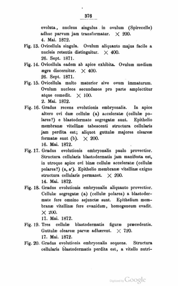
evolnta, nucleus singulus in ovulum (Spirecelle)
adhuc parvum jam transformatur. X 200.
4. Mai. 1872.
Fig. 13. Ovicellula singula. Ovulum aliquanto majus facile a
nucleis retentis distinguitur. X 400.
26. Sept. 1871.
Fig. 14. Ovicellula eadem ab apice exhibita. Ovulum medium
ægre discernitur. X 400.
26. Sept. 1871.
Fig. 15. Ovicellula multo maturior sive ovum immaturum.
Ovulum nucleos secundanos pro parte amplectitur
atque comedit. X 100.
2. Mai. 1872.
Fig. 16. Gradus recens evolutionis embryonalis. In apice
altero ovi duæ cellulæ (a) acceleratæ (cellulæ
polares?) e blastodermate segregatæ sunt. Epithelio
membranæ vitellinæ tabescenti structura cellularis
jam perdita est; aliquot guttulæ majores oleareæ
formatæ sunt (b). X 200.
16. Mai. 1872.
Fig. 17. Gradus evolutionis embryonalis paulo provectior.
Structura cellularis blastodermatis jam manifesta est,
in utroque apice ovi binæ cellulæ acceleratæ (cellulæ
polares?) (a, a’). Epithelio membranæ vitellinæ exiguo
structura cellularis permanet. X 200.
14. Mai. 1872.
Fig. 18. Gradus evolutionis embryonalis aliquanto provectior.
Cellulæ segregatæ (a) (cellulæ polares) a
blastodermate fere omnino sejunctæ sunt. Epitheiium
membranæ vitellinæ fere evanidum, homogeneum evadit.
X 200.
17. Mai. 1872.
Fig. 19. Tres cellulæ blastodermatis figuræ præcedentis.
Guttulæ oleareæ parvæ adhærent. X 720.
17. Mai. 1872.
Fig. 20. Gradus evolutionis embryonalis sequens. Structura
cellularis blastodermatis perdita est, a vitello nutri-
<< prev. page << föreg. sida << >> nästa sida >> next page >>